Posts Tagged ‘functional anatomy’


images_2

By Michael Rizk, CPT, ART  


Have you ever witnessed a relationship gone sour? The telltale signs are significant, but many  

times the root cause stems from poor communication. There is one particular area of the body  

that seems to get more press than a short‐lived Hollywood hook‐up. You’ve got it – the lumbar  

spine (LS) is likely the most injured, dysfunctional, and (supposedly) weakest link of the body.    

  In relation to the rest of the body, the LS is active in nearly every functional task performed … it  

resides at the crossroads of the body. For that reason, it is important for the rest of the body to  

communicate with the low back to let it know how important it is, how much it is needed, and  

how much it is appreciated for all it does.    

  What is meant by communication with the LS? Great question! Communication refers to the  

ability of all our joints to feed triplane motion to the LS creating triplane stability. A lack of  

triplane  mostability  (mobility  plus  stability)  can  shut  down  the  phone  lines  feeding  

proprioceptively rich information to the LS, thus creating undesirable chain reactions.  

  By design, the LS facilitates flexion and extension, allows lateral flexion, and almost inhibits  

transverse plane motion. The small amount of transverse plane motion may in fact be the most  

important motion allowing the LS to be the transverse plane transmitter of forces between the  

upper and lower extremities.   

  To simplify motion, we consider two phases: loading and unloading. Loading is the preparation  

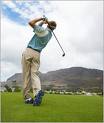
of the task and unloading is the performance of the task. Using the golf swing as an example,  

the backswing is the load and the downswing / follow through is the unload. The moment of  

time between the load and unload is what we call the transformational zone (TZ). The TZ is  

where  motion  is  decelerated  and  transformed  into  a  concentric  production  of  force.  

Understanding what happens just as we enter and exit the TZ will allow us to effectively assess  

our patients and clients.  

  I  recently  assessed  a  57‐year‐young  right‐handed  golfer  with  right  low  back  pain,  which  

occurred during the end range of his back swing just prior to transition. His approach to me was  

simple, “So I heard you can fix my back.” With a humble smile, I explained how the body works  

relative  to  the  intended  task.  I  shared  with  him  how  a  lack  of  three‐dimensional  motion  

(communication) at any segment will become excessive compensatory motion elsewhere.    

 While his assessment started with a gait evaluation, I kept in mind that gait and golf create very  

different chain reactions in the TZ.  During gait, the pelvis and trunk move opposite each other,  

and in golf, they move in the same direction. However, I immediately noticed an inability to  

load the left side of his body during gait, which was evident by a rough transition from his right  

to left foot, as well as an early heel rise on the left foot. After viewing this global glitch, I had to  

become a biomechanical detective and get more specific.  

 To  further  assess  his  left  ankle  and  left  hip,  I  had  him  perform  a  three‐dimensional  balance  

reach  matrix.  He  lacked  balance  when  I  asked  him  to  reach  in  the  frontal  and  transverse  planes  

from  his  left  foot.  To  create  stability,  I  placed  him  in  the  TRUEStretch™  in  a  backswing  posture  

and had him perform his balance reaches from this golf‐specific position. Lo and behold, he said  

“Wow,  it  feels  like  my  ankle  doesn’t  want  to  turn  that  way!”  He  continued  to  say,  “I  wonder  if  

the left ankle sprain I had playing college basketball has anything to do with this?”    

Next,  I  positioned  him  in  his  backswing  posture  to  his  threshold  of  success  (meaning  prior  to  

experiencing  any  pain  or  compensation)  and  used  my  hands  as  a  driver  to  facilitate  frontal  and  

transverse  plane  subtalar  joint  (STJ)  motions.  To  balance  his  new  mobility  with  stability,  we  

performed  lateral  and  rotational  lunges  with  three‐dimensional  arm  drivers  specific  to  the  golf  

TZ.  This  strategy  facilitates  proprioceptive  communication  between  his  left  STJ  and  right  LS.  

Within  one  week,  my  client  was  enjoying  a  pain  free  backswing  with  added  yardage  and  accuracy.  Needless‐to

say, we were both happy. Even though my client came to me with low back pain, I am led to believe his lack of frontal and  

transverse  plane  motions  at  his  left  STJ  was,  at  least,  in  part  the  CAUSE  of  his  LS  dysfunction.  If  

we  take  a  snapshot  of  the  backswing,  we  can  see  the  following:  right  trunk  rotation,  right  hip  

internal rotation, left hip external rotation, right ankle inversion, and left ankle eversion. Frontal  

plane  eversion  of  the  left  STJ  transforms  into  rotation  of  the  left  limb;  therefore,  a  lack  of  that  

motion  must  be  made  up  somewhere  else  in  the  kinetic  chain.  With  the  nominal  amount  of  LS 

rotation available there, is not much room to compensate at the LS before dysfunction and pain are experienced.  

With  an  understanding  of  Applied  Functional  Science  (AFS)  and  chain  reaction  biomechanics  we  

can  effectively  and  efficiently  keep  the  LS  healthy  and  functional  with  improved communication.  

Oh how I love function!

Get Strong! Stay Strong!

Chris


images_3

 By Barbara Fuller PT, FAFS

The late-seventyish woman looked at me with a bit of skepticism mixed with a little fear. After I introduced myself to her she said, “Before we start, I want you to know that I would like for you to address my low back pain, but I don’t want you to mess with my hip.” When I urged her, she further explained that she’d had problems with her right hip for a long time, with x-rays revealing severe arthritis and necrosis of the femoral head. She had been to an orthopedist who told her to come back for a total hip replacement when she could not stand the pain anymore. She was not ready for that. Her back pain was relatively new, but it was making her even more miserable, as well as making her unable to do the yard work she loved doing or taking a walk. Most telling was the account of her last experience in physical therapy when her hip was forced passively beyond what she could tolerate, thus resulting in increased pain. Needless-to-say, she never went back. Before I even touched this patient, or put her through any functional movement assessments, I felt a strong need to educate her about the hip – especially its relationship to the lumbar spine. I told her that the hip is the low back’s best friend. I explained how a normally functioning hip, one that moves well and is strong in all three planes of motion, allows the low back to function normally by protecting it from excessive motion and wear and tear. I demonstrated how a hip, with a decent amount of extension, propels the body forward, but without that extension, the task falls to the low back causing all sorts of problems. I told her that her hip had let her low back down and was probably the cause of her low back pain. All that being said, I then told her that I really felt that I needed to at least take a look at the hip or I would be doing her a disservice. I also promised her that I would not do anything she did not agree to me doing and that I would be very gentle. She agreed. In addition to the hip being quite friendly, the hip is BIGGEST of the BIG rocks in our body! Big rocks are the areas of the human body that provide a foundation for normal mobility and stability (mostability). But if they are not functioning well, then they become the culprits of pain and dysfunction in joints above and below. For the hip, that includes its closest neighbors – the lumbar spine and the knee – but it can also include more distant neighbors like the opposite shoulder or elbow. Also, the hip is our power source. We can understand this when we consider the powerful muscles of the back and front butts, and their core connections, including the pelvic floor (with connection through the hip adductors and rotators) and the thoracic diaphragm (with connection through the abdominals to the thoracic spine and rib cage). It is through hip extension that we can most effectively load our abdominals. As promised, I was gentle with my patient and started her functional assessment with neutral standing (XXX) while hanging on to a counter top with both hands. She was limited bilaterally in lateral pelvic glides and rotations. Most significant, though, was her lack of right hip extension in a left stride stance (LXX). She also demonstrated to me a successful short squat – something that applied to her goal of continuing with yard work. Her home-workable exercise program (remember the test becomes the exercise and the exercise becomes the test) became XXX with right and left lateral glides and rotations of the pelvis for a warm-up, which was then followed by LXX anterior and posterior glides. After these, I asked her to do some of her short squats (start with success). On her next visit she beamed and said, “I’ve had very little pain since my first visit!” She progressed beautifully through her physical therapy. Although she did not get full mostability back in her right hip, she gained just enough that on the day when she was able to complete a common lunge matrix (anterior lunge, same side lateral lunge, and same side rotational lunge) – no hands – she looked at me and said, “I feel so empowered!” Wow! I did not heal this patient, but provided an environment in which her body could improve. By giving her complete control, her fears were addressed. By educating her about the truth of the hip, her skepticism was addressed. And by giving her a home-workable program, she was empowered … and so was her hip! This is the beauty of Applied Functional Science.

Get Strong! Stay Strong! (and don’t forget to be functional!)

Chris

images_47 

Not that it could ever happen, but if given the opportunity to defend a body part in a court of law, without question I would choose to defend the lumbar spine. With mind boggling statistics such as 80% of all human beings will experience low back pain at some time during their life and that the cost of low back injury is estimated at being $100 Billion each year, there is no wonder why the lumbar spine has been the subject of much maligning and is often referred to as “a bad back”. Despite those daunting statistics, I would argue that not only is the lumbar spine ‘not guilty’ but is in fact most often a victim. The true culprit in low back dysfunction is most often a problem with a link or two in the biomechanical chain somewhere other than the low back. 

     The Lumbar Spine is located at ‘the crossroads of the body’. It lives where everything happens. It is a five segment stack at the base of the spine designed with the vertebral body and intervertebral disc in the anterior segment and bony protrusions in the posterior including the facet joints to either side. In a typical and healthy back the Lumbar spine has between 40-43 degrees of motion into flexion, 30-40 degrees of flexion into extension, 20 degrees of frontal plane side-bending in each direction and only 5 degrees of transverse plane rotation to each side. Initial examination may lead one to conclude that with such limited frontal and transverse plane motion, there is no wonder that the lumbar spine is often breaking down. Some might say ‘This is a terrible design flaw’. However, knowledge of chain reaction biomechanics and Applied Functional Science allow for deeper understanding.

     When fed proper proprioceptive input and when surrounded by ‘friends’ who are functioning properly, the lumbar spine is perfectly equipped to carry out its roles as a shock absorber and force transmitter. The lumbar spine enjoys enough motion to allow the rest of the system to be turned on, facilitating functional strength and motion requirements elsewhere. However, when the lumbar spine is let down by its friends it tries to help the situation by picking up the “slack”. Unfortunately to do so it must work beyond it’s capabilities to try and get the functional job done. That is when the problems start.

images_48

     It is a plausible contention that when the low back starts to hurt, look everywhere else and you’ll find the reason why. For example, say the lumbar spine has just distal to it, two under-nourished hips (please read Joe Przytula’s fantastic take on the importance of the hip in upright 3d function from the June newsletter!). These hips are typically allotted a generous amount of transverse plane mostability (right amount of motion, right amount of stability). However, when they are not ‘properly fed’ the tendency is to seek stability rather than mobility and transverse plane motion at the hips is lost in function. Who then steps in to help? The under-equipped lumbar spine. With the possibility of only 5 degrees of available motion, wear and tear is going to start quickly especially if the person is active despite their hip restrictions. 

     It would be easy if the hips were the only friends capable of letting down the lumbar spine. Typically, however, uncovering the underlying cause behind low back dysfunction is more challenging. Because it is located at the crossroads, limitation in virtually any other biomechanical segment in any plane of motion has the potential to feed abnormal and damaging forces through the lumbar spine. Hopefully your lumbar spine is surrounded by good friends who feed it appropriate forces. If not, and it has been referred to as ‘a bad back’… beware! We may team up in a defamation suit!   —Andrea Wasylow PT

Get Strong Stay Strong!

Chris


images_46 

A few weeks ago, my six-year-old nephew was sleeping over at our house. I was awaken in the middle of the night by him exclaiming, “Uncle Joe, there’s a monster underneath my bed!” I went into his room and said, “Be nice to him and he’ll be your best friend. Give him a pop tart or something.” Okay, maybe I’m not the best uncle, but please read on.

The hip is the like a monster underneath the bed, minus a few dust balls and dirty magazines. If it’s working correctly, it can be your best friend. It can assist far away joints like the shoulder or ankle. Strength Coach Vern Gambetta called it, “The engine that drives the body.” No wonder, it has 17 of the thickest, longest muscles of the body directly attached to it. But do not look for them. They are superficially hidden by that big mattress we call the gluteus maximus. A few years back, researchers Porterfield & DeRosa discovered the monster even has tentacles! Well, sort of – we call it fascia. It functionally links the hips with pretty much the entire body.

images_2

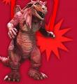
This monster is tough all right. It has a deep suction cup of an acetabulum, with a head of the femur as round as Mini-Me’s head, and a thick synovial joint capsule to seal the deal. How does the hip stack up to other monsters, say Godzilla (he just spit fire)? The hip’s secret power is its contribution to three-dimensional loading (force reduction) and unloading (force production). Let’s use the ACL ligament of the knee as an example. Traditional rehab protocols have emphasized the quadriceps and hamstrings. However, physical therapist and biomechanist Daniel Cipriani makes the point that these muscles only become protective as the knee flexion angle approaches 90 degrees. But now look upstairs at the hips gluteus complex. By way of its multidirectional, multiplane orientation on the femur, it is well designed to control the 3D motion of the knee with the most critical being internal rotation, adduction, and flexion.

images_3

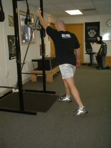
Let’s follow those tentacles up the kinetic chain to the shoulder. Can they protect the shoulder anterior instability that creates rotator cuff issues? You bet! Now we’ll call on the infamous “front butt,” including the iliopsoas, abdominals, adductors, rectus, etc. Try it yourself: Stand in a left stride stance with your right arm horizontally abducted at shoulder height with the elbow flexed. Feel the tug at the front of hips? This means they’re locked and loaded to explode, and so are the abs by way of chain reaction between the hip and shoulder. Now turn the front butt off by sitting on it. Do the same arm reach. Feel the difference?

We make the monster happy when we feed it. No, not with pop tarts. Hips love ground, gravity, and momentum. They prefer lunges, squats, and step-ups. Adding some arm reaches in with the mix is like whip cream on top. They love variety in the form of direction, plane, speed, and load changes. However, be cautious of feeding the hips with empty calories. Many exercises performed in the prone, side lying, or supine positions are what Gary Gray refers to as “isolated isolation.” They turn off the hip’s phone lines (proprioceptors) to the rest of the body and unhook those fascia links. They should be used sparingly. Dormant daily living does not nourish the hip. Sitting and activities that require prolonged static standing promotes injurious capsular patterns.  Interrupting these patterns with frequent “snacks” helps reconnect those lines.

By Joe Przytula ATC

Get Strong! Stay Strong! (Functionally)

Chris

In the previous hip flexor post we talked about how the tight hip flexor inhibits the gluteus maximus and increased lumbar extension and hamstring activity to compensate.  There are also several other compensatory problems that occur from the tight hip flexor.  Remember that most people have tight hip flexors due to sitting, driving, sleeping in the fetal position, inactivity and repetitive patterns.  So, if the hip flexor is tight it could lead to patellar tendon/knee pain, and plantar fasciitis.

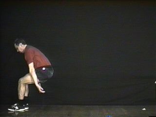
Lets look at jumping.  Jumping requires an eccentric load (hip/knee/ankle flexion and internal rotation) followed by a concentric contraction (hip/knee/ankle extension and external rotation).  In the take off phase if the hip flexor is tight it inhibits the gluteus thereby decreasing its ability to generate force.  This leads to poor jumping ability.  It also limits the amount of hip extension that can occur therefore the knees will hyperextend as a compensation for the lack of extension at the hip.  The hyperextension and the increased quad force cause the patella to be compressed and inferiorly tilt more irritating the tendon and fat pad leading to knee pain and tendonitis.  

Upon landing the gluteus cant control the forces and the femur excessively internally rotates and adducts causing increased stress to the med knee. ( The gluteus maximus eccentrically controls femoral internal rotation during loading).  This can be one of the causes of poor jumping mechanics which has been seen especially in females and correlated too increased ACL injury risk.

If the hip flexor is tight it will decrease extension of the hip and therefore decrease stride length.  This can lead to increased extension of the low back which over time can cause low back pain or as a result cause the heel to come off the ground early in the trail leg of gait.  This results in the trail leg diminishing its eccentric loading and therefore decreasing its ability to push off efficiently.  This ultimately creates more stress on the front leg as the muscles have to work harder to re – supinate the foot.  The lead leg in gait relys on the push off and successive swing of the rear leg to re-supinate and unload.   With the trail leg heel coming off the ground early an a-propopulsive gait results.  While this is a mild stress that wont cause problems in one step, the microtrauma and/or fatigue over time (thousands of steps in a day, 7 days a week and 4 weeks in a month etc.) eventually leads to the tissue unable to tolerate the stress and bang, pain occurs in this case plantar fasciitis.  So, a tight hip flexor on one side can lead to plantar fasciitis on the opposite side.  The beauty and challenge on understanding function, it never gets old!

Get Strong! Stay Strong!

Chris

Most anatomy classes teach that the hamstrings flex the knee.  Based on this many people train and strengthen the hamstrings using a leg curl machine either sitting or laying on their stomach.  If we stop and look at the body function in various activity you will see that rarely ever are people sitting or laying prone and flexing their knees.  Even in walking or running when you see the knee flexing it is a passive event caused by momentum.

Lets first look at the anatomy.  In general. the hamstring attaches proximally to the ischial tuberosity (the bone you sit on) and runs down the back of your leg to attach distally to the medial and lateral tibiia (lower leg bone).  Next lets consider the “true” function of this muscle.  Using gait or a lunge as an example, knowing what the bones are doing (in all 3 planes) and knowing where a muscle attaches prox. and distally will allow you to see and figure out function of any muscle.  Remember that in the loading phase (eccentric muscle activity) the tibia advances forward and internally rotates and the pelvis/hip flexes, adducts and internally rotates (see when the foot hits ground and “barking hip posts for review).  So with that in mind, as the foot swings forward the hamstring muscle eccentrically contract to decellerate the leg to prepare it for heel strike.  When the foot hits the ground and begins to load the hamstrings eccentrically contract to control hip flexion in the sagittal plane (so we dont fall on our face).  They eccentrically control tibial internal rotation in the transverse plane and help control hip adduction (in single limb stance with the glut med / min) in the frontal plane.  Distally, think of the hamstrings like reigns of a horse controlling and acellerating tibial rotation.  Once the foot gets to late midstance we have completed the loading phase and the muscles now concentrically contract to propel us forward. The hamstring concentrically contracts to extend the hip and externally rotate the tibia.  How well the hamstring (or any muscle) concentrically unloads or explodes contracts depends on how efficiently it eccentrically loads.  Based on the biomechanics and “functional” anatomy one can see how doing leg curls will not prepare you to walk, run or lunge.  When considering exercise for function or performance it is important to consider how the muscle or joints respond to gravity, ground reaction force (GRF) and momentum.  Gary Gray refers to this as training in the context in which you will actually utilize the muscle and joints in relation to GRF, gravity and momentum.


Some examples (above) of functional hamstring exercise consist of single leg (SL) squats w/ reaches, lunges with reaches, isom. SL squat w/ alternating cross reaches modified deadlifts, and SL mod. deadlifts.

Get Strong! Stay Strong!

Chris

OK, since I enjoy talking about function and biomechanics lets talk about how to turn on your butt!  Thought that might get someones attention.  Any way, the Gluteal/hip/ butt muscles are the “powerhouse” muscles.  We call them the big house or the cannon.  The first thing to do before “shooting” the cannon is to “load” the cannon.  In function this simply means you need to efficiently load (eccentrically)the gluteus muscles in order to effectively unload or more forcefully create a concentric contraction.  During the loading or pronation phase ( front foot hits the ground in lunge or gait)

   the function of the gluteus muscles (max, min and med) is to eccentrically control hip flexion in the sagittal plane, hip adduction in the frontal plane and femoral/hip internal rotation.  If you look at an anatomy picture of the gluteus maximus you will notice that the fiber orientation is primarily in the transverse plane.  This baby was designed for femoral/hip rotation (a significant component of lower extremity pronation)

The gluteus attaches proximally to the saccrum and the illiac crest and distally to the greater trochanter and blends with the tensor fascia latae (TFL) to form the illiotibial band.  The ITB attaches distally to the anterior/lateral aspect of the tibia.

So, for example, when you are walking and your foot hits the ground and begins to pronate, the calcaneus everts causing the tibia to advance forward and internally rotate (see “When Foot Hits Ground” post).  This creates a quick “pull” on the ITB, which creates a quick stretch on the gluteus thereby stimulating the butt to contract.  This is further accentuated by the femoral internal rotation that also eccentrically lengthens the gluteus maximus.  Remember that neurologically a muscle responds to a quick stretch (eccentric load) by concentrically contracting. Therefore, if one has limited calcaneal eversion (after ankle injury or immobilization) they will not be able to effectively and efficiently turn on the butt which leads to compensation and further problems.  There in lies the beauty of function!


Get Strong! Stay Strong!

Chris
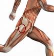
Posterior Tibialis: Attaches proximally to post aspect of tibia and distally attaches to almost everything under the foot (cuboid, navicular, cuneiforms).  During pronation (loading phase) it eccentrically controls tibial advancement in the sagittal plane(SP), tibial internal rotation in the transverse plane(TP) and eccentrically controls lateral to medial loading of the foot in the frontal plane(FP).

Soleus: Attaches proximally to post surface of tibia and distally forms achilles tendon to attach slightly medial on the calcaneus.  During the loading phase it eccentrically controls tibial advancement in the sagittal plane, tibial internal rotation in the transverse plane and calcaneal eversion in the frontal plane.

In most anatomy classes it is taught that the function of the posterior tibialis. is to plantar flex (point) and invert the foot and the soleus plantar flexes the foot.  Unfortunately, when your foot is on the ground this does not and cannot happen as was explained above.  After an injury or surgery many people walk on a bent knee.  Assuming they have full passive extension, the problem is a posterior tibialis and soleus that are not functioning properly.  When the foot hits the ground and begins to pronate, the soleus is ecc. controlling calcaneal eversion (FP), the post. tib. and soleus are ecc. controlling tibial advancement(SP) and internal rotation(TP) and the tib. post. is controlling the lateral to medial loading of the foot.  This all occurs through late mid stance of gait (normal pronation).  Then the foot/leg begin to supinate (unload) in order to lock up the joints to push off on a rigid lever.  The soleus inverts the calcaneus while the post. tib. and soleus concentrically “pull” back on the tibia and externally rotate it.  Meanwhile momentum is carrying the rest of the body “over the top” and thats what creates knee extension during gait.  It really has nothing to do with the quad, which is taught in school to extend the knee.  School anatomy really didnt prepare me or others to treat movement dysfunction and injury in the real world.  Thank goodness I was fortunate to meet Gary Gray early in my career.  He really opened my eyes to true “functional anatomy.”  For that I am grateful.

It is truly a never ending journey.

Get Strong! Stay Strong!

Chris
Many people are plagued by low back pain (LBP). It ranges from severe pain and disability to general achiness and stiffness. Many (not all) of today’s problems can be linked to one simple fact—we sit too much! In fact, technology has created a society that you don’t even have to move to be part of. This leads to muscle and joint stiffness, decreased flexibility, poor posture and deconditioning. In particular the hip flexors, hamstrings, calves and chest get tight while the upper and lower back get rounded. This creates muscle imbalances and causes specific muscles to be “turned off” via reciprocal inhibition. Then, when and if we decide to do something, ouch! Pain develops. Sometimes the pain can be linked to a specific incident, but many times it gradually occurs with no seemingly apparent reason. The latter is where the compensatory effects of posture and deconditioning (becoming more cmmon in todays society) come into play. For example, when you sit a lot the hip flexors are in a shortened position and over time will get tight. Upon standing the hip extends. If the hip flexor is tight the hip won’t be able to extend through its full range and can place more stress on the lumbar spine as it tries to extend more to make up for the lack of motion its supposed to get in the hip. This not only occurs with standing but with every step you take. Over time this can increase the wear and tear on the spine and be setting you up for back pain, in addition to a number of other problems. There is good news. Many of the problems associated with LBP can be significantly reduced and prevented with the help of a knowledgeable professional. Understanding “functional” anatomy and biomechanics allows the professional to evaluate, identify altered movement patterns, weakness and inflexibity in order to develop a custom program that will get you moving in the right direction.  Personally, one of my favorite books on low back is Stuart McGills Ultimate Back Fitness and Performance.

Get Strong!Stay Strong!

Chris